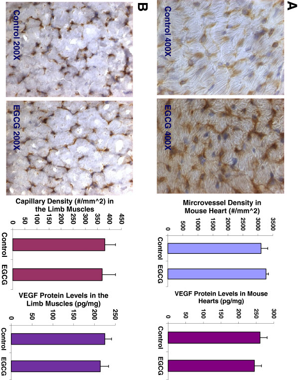
Figure 5.
EGCG treatment did not affect the capillary density (3270 ± 162 vs. 3103 ± 226 #/mm^2; n = 8; P = 0.5215), and VEGF expression (261 ± 22 vs. 245 ± 19 pg/mg; n = 8; P = 0.4517) in the mouse heart, compared to the control group (Panel A), respectively. There was no significant difference in the capillary density (370 ± 55 vs. 381 ± 44 #/mm^2; n = 8; P = 0.5401), and VEGF expression (225 ± 16 vs. 214 ± 20 pg/mg; n = 8; P = 0.7825) in the limb skeletal muscles between the EGCG-treated mice and the control mice (Panel B), respectively. The digital images show CD31 immunohistochemistry staining in OCT-embedded cryosections of the heart (Panel A) and the limb muscle (Panel B) of control mouse and EGCG-treated mouse, respectively.

