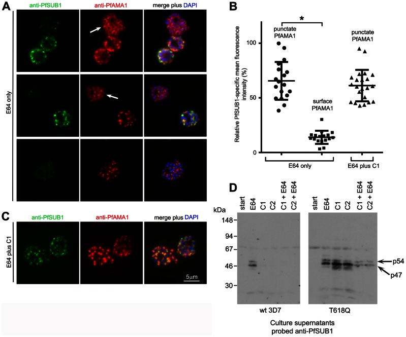
Figure 3. PfPKG inhibitors block discharge of exonemes and micronemes.
(A) IFA of wt 3D7 parasites allowed to develop beyond the point of egress in the presence of E64 only (50 µM). Top two rows: segmented schizonts displaying a merozoite surface localisation of PfAMA1 (arrowed; ∼45% of the schizont population) as a result of its discharge from micronemes, exhibit a weak PfSUB1 signal compared to less mature schizonts in which both the PfAMA1 and PfSUB1 signals remain punctate. The bottom row of images in (A) shows a schizont in which the PfAMA1 signal is intermediate between punctate and merozoite surface. (B) Surface translocation of PfAMA1 is associated with loss or redistribution of the PfSUB1-specific IFA signal. The PfSUB1-specific mean fluorescence intensity in individual schizonts relative to that of the PfAMA1-specific signal was acquired as described in Materials and Methods. Individual relative fluorescence intensity values (at least 17 schizonts per group) are shown plotted, with mean and SD indicated. Within the parasites treated with E64 only, values were significantly lower for the schizonts displaying the merozoite surface PfAMA1 phenotype (Student's unpaired t-test, two-tailed P value<0.0001). There was no significant difference between the E64-treated and E64 plus C1-treated values for schizonts with the punctate PfAMA1 phenotype (see panel C). (C) IFA of wt 3D7 parasites allowed to develop beyond the point of egress in the presence of both E64 and C1. No discharge or relocalisation of PfSUB1 or PfAMA1 was evident in counts of >5,000 schizonts from a total of 3 independent experiments. Identical results were obtained with E64 plus C2 (not shown). (D) Western blot of culture supernatants from synchronous wt 3D7 or PfPKGT618Q schizonts allowed to develop beyond the point of egress in the presence of E64 only (50 µM), C1 only (2.5 µM), C2 only (1.5 µM), or indicated combinations. Cultures were sampled immediately (start) or after incubation for 4 h. The p54 and p47 forms of PfSUB1 usually observed following its maturation are arrowed. The relatively low levels of PfSUB1 in supernatants of the PfPKGT618Q parasites in the presence of C1 or C2 plus E64 are likely due to the block in egress mediated by E64. See also Figure S3 in Text S1.

