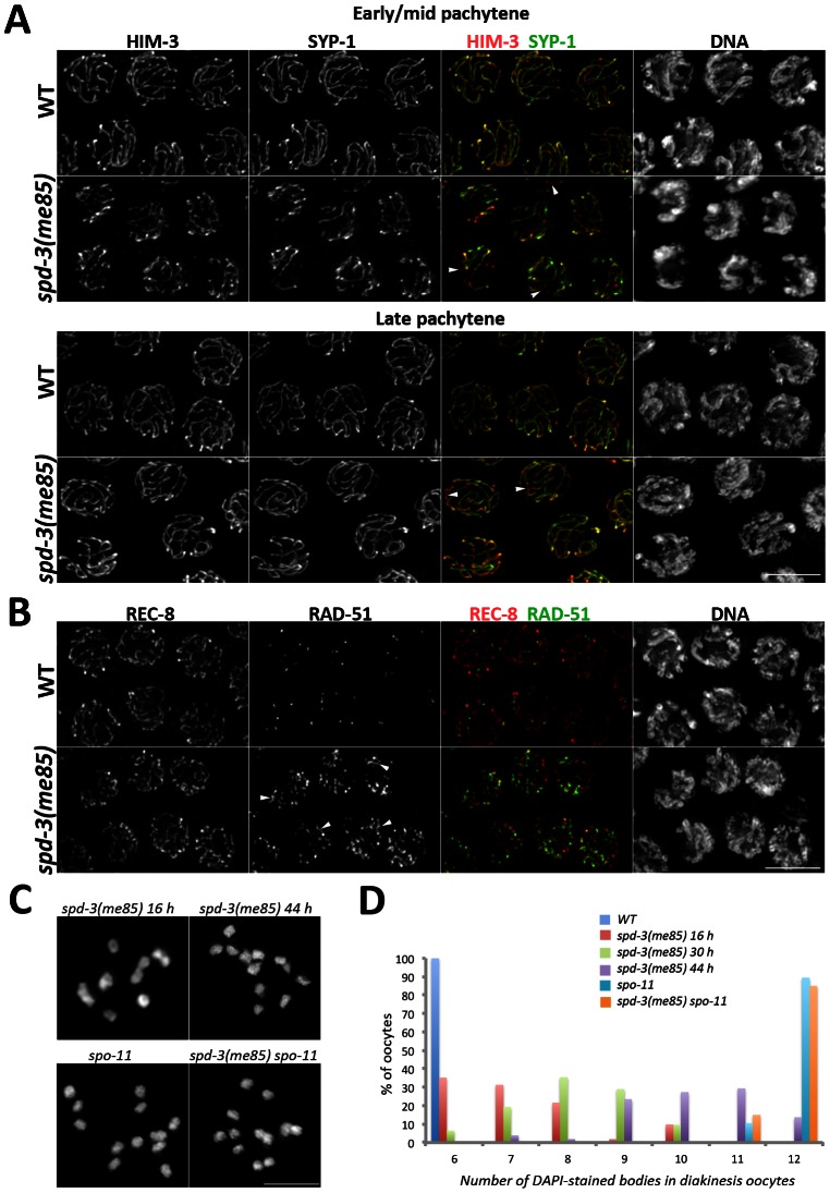
Figure 2. SC assembly and recombination in spd-3(me85) mutants.
(A) Projections of pachytene nuclei stained with anti-HIM-3 and anti-SYP-1 antibodies and counterstained with DAPI. Arrowheads point to unsynapsed regions. (B) Projections of pachytene nuclei stained with anti-REC-8 and anti-RAD-51 antibodies and counterstained with DAPI. Arrowheads point to long stretches of RAD-51 signals present in the spd-3(me85) mutant. (C and D) Projections of individual diakinesis oocytes of the indicated genotype stained with DAPI (C) and quantification of the number of DAPI-stained bodies present in diakinesis oocytes of the indicated genotypes and ages (D). 6 DAPI-stained bodies corresponds to 6 bivalents (wild-type oocytes), while 12 corresponds to 12 univalents (no crossovers) and 7 to 11 indicate a mixture of bivalents and univalents. The number of diakinesis oocytes analyzed per genotype were: spd-3(me85) 16 hours (52 oocytes), spd-3(me85) 30 hours (31 oocytes), spd-3(me85) 44 hours (51 oocytes), wild-type control 16 hours (52 oocytes), spo-11 16 hours (30 oocytes), spd-3(me85) spo-11 16 hours (40 oocytes). A two-tailed Mann-Whitney test shows that the difference between any two of the analyzed genotypes is highly significant (p<0.001), apart from the comparison between spo-11 and spd-3(me85); spo-11 which is not different (p = 0.7). Scale bar = 5 µm in all panels.

