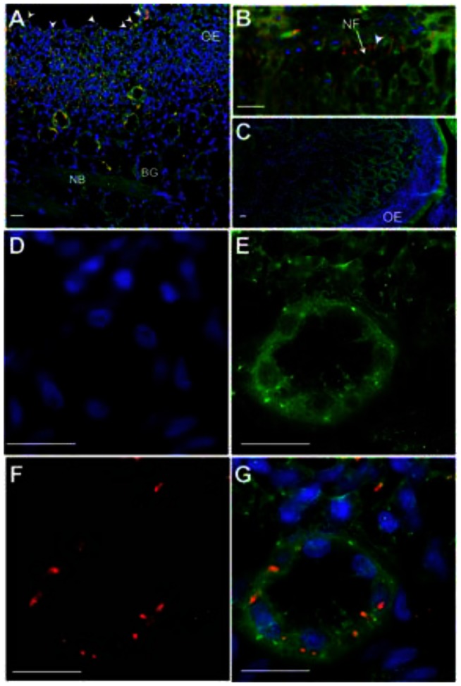
Figure 7. Tracking lyophilized prion inocula in the nasal cavity of deer.
Highly enriched, fluorescently labeled prions were mixed with Mte, lyophilized, pulverized and puffed into the nasal cavity. (A) After 45 minutes, florescent prion aggregates (red) can be seen on (white arrowheads) and within the olfactory epithelium (OE) of the nasal turbinates. Tissue sections are counterstained with DiOC18 fluorescent membrane dye (green) and the nuclear stain DAPI (blue). A small proportion of prions can be seen near serous cells of the Bowman's glands (BG) in the lamina propria. (B) By 60 min, significant amount of prions were found in the lamina propria, with some aggregates (arrowhead) associated with nerve fibers (NF) emanating from the OE. (C) We detected no red signal from negative control sections from mock-inoculated deer. (D–G) Higher magnification of a Bowman's gland stained with DAPI (D), DiOC18 (E) and decorated with prions (F) that appear to localize on serous cells (G). NB, nerve bundle; scale bar, 20 µm.

