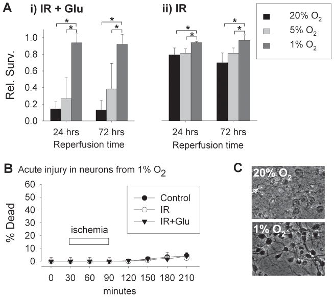
Figure 2. Protective effects of hypoxic adaptation against ischemia-reperfusion injury.
A) Relative survival of cortical neurons 24 and 72 hours following 60 min simulated ischemia-reperfusion with 30 μM glutamate (i) and simulated ischemia without added glutamate (ii). Neurons were maintained from time of plating in 20%, 5%, or 1% O2 tensions to DIV 9, when they were subjected to simulated ischemia. Following ischemia, all neurons were returned to conventional 20% O2 conditions for 24 to 72 hours. Neurons adapted to 1% O2 were strongly protected from reperfusion injury (n = 4 and 3, at 24 and 72 hours, respectively; mean ± S.D.; *p<0.05 in two-way ANOVA testing followed by Student-Newman-Keuls post-hoc testing). B) Acute neuronal death (percentage of cells taking up propidium fluorescence) in cells exposed to oxygenated, glucose-containing saline (control) or to simulated ischemia-reperfusion, without or with added glutamate (IR, or IR+ Glu). C) In neurons adapted to conventional 20% O2 conditions, widespread dramatic swelling and acute necrosis resulted within 2 hrs of reperfusion following simulated ischemia (upper panel; arrow indicates an example of an acutely swollen neuron with nuclear propidium fluorescence). In neurons adapted to 1% O2 (lower panel), no measurable cell death occurred (p = 0.66 for comparison of O2 levels, two-way ANOVA).

