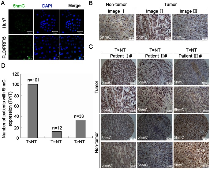
Figure 1. The level of 5 hmC was decreased in human HCC.
(A) Detection of 5 hmC in HCC cells (Huh7 and PLC/PRF/5) by immunofluorescent staining. The representative photomicrographs were presented. (B) Detection of 5 hmC in the paraffin-embedded formalin–fixed HCC tissues by immunohistochemistry. Representative photomicrographs were presented. (C and D) The level of 5 hmC was examined in a tissuearray containing 146 paraffin-embedded formalin–fixed HCC tissues and paired non-HCC counterparts by immunohistochemistry. The representative photomicrographs of 5 hmC level with high, equal and low in HCC tumors, as compared with non-tumor tissues, were shown (C). The altered level of 5 hmC between HCC and non-tumor tissues in 146 HCC patients were summarized (D) (n = number of cases).

