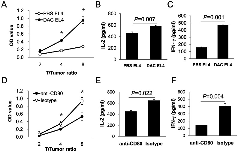
Figure 8. Role of DAC induced CD80 expression in stimulation of T cells.
Six million irradiated EL4 cells were injected i.p. into each BALB/c mouse twice with a week interval between injections. Spleen and lymph node cells from EL4 immunized BALB/C mice were co-cultured with DAC-treated or PBS-treated EL4 cells for 6 days. (A) T cell proliferation was assessed using Cell Counting Kit-8 (CCK8, Dojindo, Kumomoto, Japan) on day 6. IL-2 (B) and IFN-γ (C) production in the culture supernatants were detected by ELISA. For CD80 blocking, spleen and lymph node cells from EL4-immunized mice were co-cultured with DAC-treated EL4 cells in the presence of 5 µg/ml anti-CD80 antibody or an isotype matched control antibody. (D) T cell proliferation was assessed using the CCK8 assay. Supernatants of co-culture were harvested 48 hours later for detection of IL-2 (E) and IFN-γ (F) by ELISA. Student’s t-test was used for statistical analysis. Data shown are representative of three experiments with similar results.

