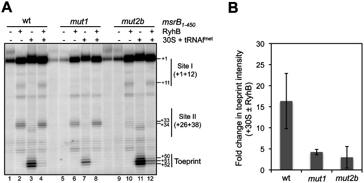
Figure 6. RyhB binding at Site I and Site II blocks ribosome binding to the msrB translation initiation region.
(A) An autoradiogram of a toeprint analysis is shown (for details, see Materials and Methods, and Results). msrB1–450 wild type and variants (mut1 and mut2b) were used as a template (5 nM) in the cDNA extension experiment. Lanes (1, 5, 9): extension with no other component added; lanes (2, 6, 10): extension with RyhB alone (2.5 μM); in lanes (3, 7, 11): extension with 30S subunit (+initiator tRNAfmet) alone (500 nM) ; lanes (4, 8, 12): cDNA extension with 30S subunits (500 nM) along with RyhB (2.5 μM). Thin vertical lines indicate nucleotides involved in Site I and Site II. The transcription start of msrB is referred to as the position + 1. The 30S subunit-induced reverse-transcriptase (RT) toeprint is indicated at positions +50 to +52. Other indicated positions are numbered accordingly. (B) Quantification of band intensity was performed by using Image J software and expressed in terms of fold change in toeprint intensity. Fold change represents decrease in toeprint intensity obtained when comparing the two conditions, with 30S ribosomal subunits alone and with 30S ribosomal subunits along with RyhB. Standard error of two independent experiments is shown.

