
Figure 3.
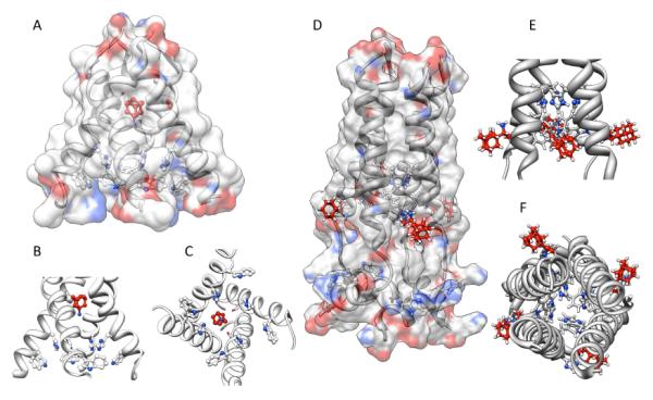
Binding modes of aminoadamantanes to Influenza A M2 model peptides. Panel A – C: Amantadine binding inside the pore of M2 transmembrane domain (X-ray diffraction data, pdb code 3c9j). Panel D – F: Rimantadine binding from the outside of the M2 model peptide (representative NMR data, pdf code 2rlf). The drug molecules are shown in red, His and Trp residues in the M2 model peptides are depictes as ball-and-stick models. See text for a discussion.