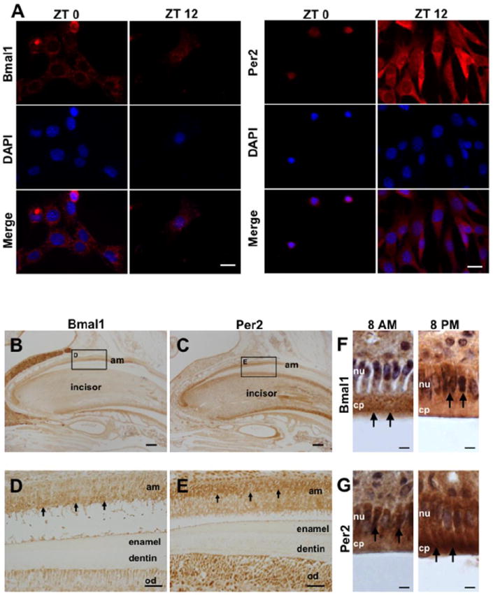
Fig. 4.

Cellular localization of clock proteins in HAT-7 during a 24h period. A) Immunocytofluorescence staining reveals that BMAL1 proteins are mostly localized in the cytoplasm at ZT0 and then shift into the nucleus at ZT12 (left). In contrast, PER2 is localized in the cell nucleus at ZT0 and then it shifts to the cytoplasm at ZT12 (right). B) IHC results show that BMAL1 (C) and PER2 (D) are expressed in ameloblasts (black arrows). On serial sections, BMAL1 is mostly expressed in the cytoplasm (D), and PER2 is localized mainly in the nucleus (E). Most of BMAL1 proteins are localized in the cytoplasm at 8 am and then in the nucleus of ameloblasts at 8 pm (F). PER2 proteins are found in the nucleus at 8 am and in the cytoplasm of ameloblasts at 8 pm. Am, ameloblast; od, odontoblast; nu, nucleus; cp, cytoplasm. Scale bars = 20 μm in A, 200 μm in B and C, 40 μm in D and E, 10 μm in F and G.
