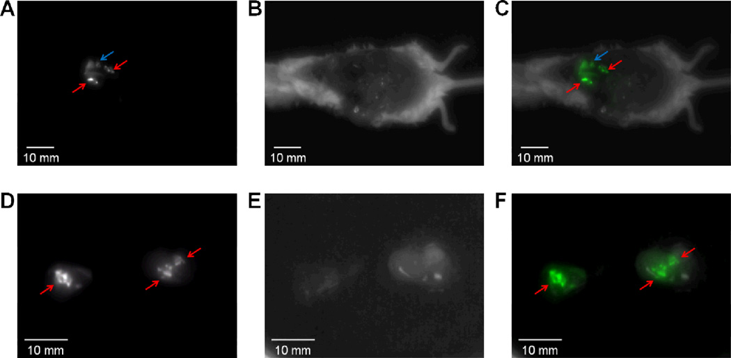
Figure 2.
Detection of diffuse satellite lesions with fluorescence goggle and ICG in Group 1 mice. Intraoperative (A) NIR fluorescence image, (B) reflectance image and (C) merged image of (A) and (B) of a mouse 48 h post-injection of ICG are shown. Fluorescence goggle detects scattered metastases and small tumor deposits in the liver intraoperatively, which are not obvious to naked eye. Ex vivo (D) NIR fluorescence image, (E) reflectance image and (F) merged image of (D) and (E) are shown. NIR fluorescence is pseudo-colored in green in panels (C) and (F). Red arrows indicate the metastasis, and blue arrows indicate the liver.

