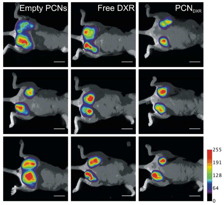
Figure 6.
Non-invasive fluorescent imaging of nude mice with mCherry-labeled MDA-MB-231 mammary tumors treated with empty PCNs, free DXR, or PCNDXR (scale bar = 1 cm). mCherry fluorescence is pseudo-colored and overlaid over bright-field images. All members in each group are shown (n = 3 mice and 6 tumors).

