
Fig. 1.
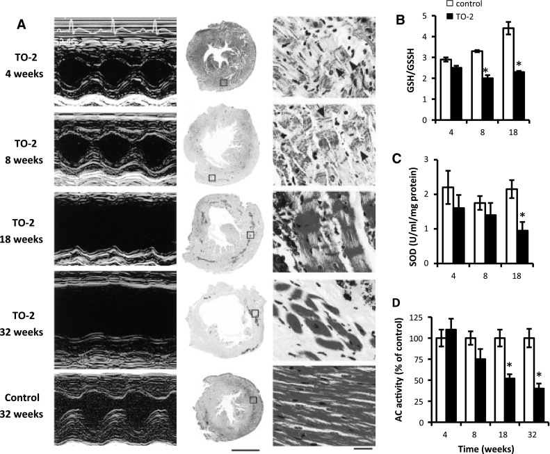
Serial changes in left ventricular (LV) function and extent of oxidative stress in TO-2 and control hamsters. A Representative echocardiograms and representative light photomicrographs of LV cross-sections stained with hematoxylin–eosin. B–D Glutathione redox ratio (GSH/GSSG; oxidized glutathione/reduced glutathione) (B), superoxide dismutase (SOD) enzyme activity (C), and adenylyl cyclase (AC) activity (D) in the LV myocardium. Data are mean ± standard error of the mean (SEM) of 8 animals in each experiment. *P < 0.05 versus age-matched controls