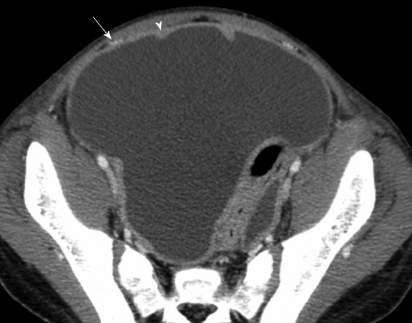
Figure 7.

Computed tomography with intravenous contrast. Thirteen years old with rhabdomyosarcoma. The anterior paravesicular spaces are well defined with ascites. The lateral umbilical fold (containing the inferior epigastric vessels, arrow) divides the lateral and medial inguinal fossa. In between the medial umbilical fold (containing the obliterated umbilical vein, arrowhead) is the supravesicular fossa.
