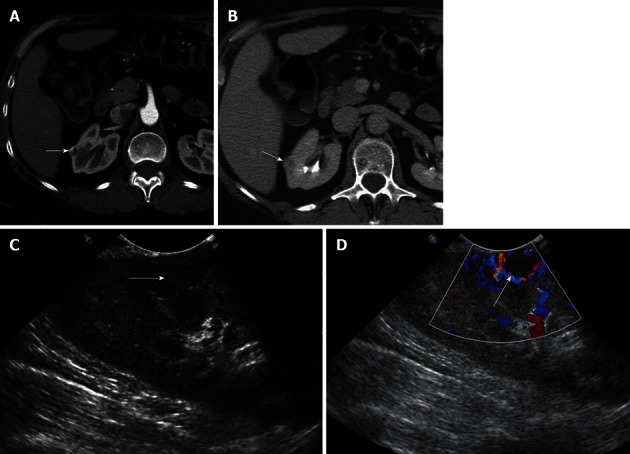
Figure 4.

A 51-year-old female with incidentally detected small solid mass in the right kidney, compatible with a renal cell carcinoma. A, B: Axial contrast-enhanced images of the right kidney show a 1.2 cm hypervascular mass in the midportion of the right kidney, concerning for a small renal cell carcinoma; C: Longitudinal Intraoperative ultrasound image localizes the small solid mass in the midportion of the right kidney anteriorly; D: Intraoperative ultrasound with Doppler interrogation shows prominent vessels at the margin of the lesion. Intraoperative ultrasound is an invaluable resource to localize small solid renal lesions during partial nephrectomy, ensuring that negative margins are achieved, while preserving the normal renal parenchyma.
