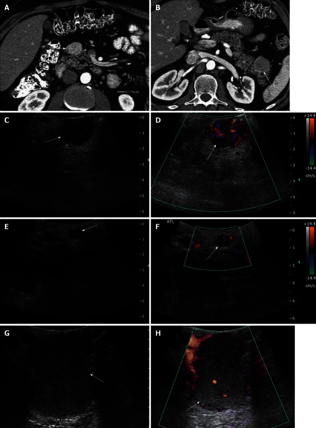
Figure 8.

A 46-year-old male with multiple endocrine neoplasia type I syndrome and pancreatic neuroendocrine tumor. A, B: Axial contrast-enhanced computed tomography (CT) images show hypervascular masses in the head and tail of the pancreas, consistent with neuroendocrine tumors; C: Intraoperative ultrasound image shows a well-defined solid mass in the head of the pancreas, consistent with the neuroendocrine tumor on CT; D: Doppler interrogation reveals increased vascularity within this mass; E, F: Intraoperative grayscale and color Doppler ultrasound images detect a 5 mm solid mass with internal vascularity in the head of the pancreas, consistent with a small neuroendocrine tumor. This lesion was not identified on the CT examination; G, H: Intraoperative grayscale and color Doppler ultrasound reveals the large dominant mass in the tail of the pancreas, consistent with a neuroendocrine tumor.
