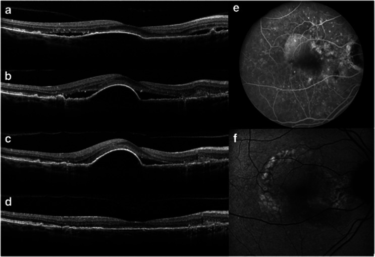
Figure 2.
Images from Case 2: a 55-year-old Caucasian female with neovascular age-related macular degeneration in her right eye. (a–d) Images from Heidelberg SPECTRALIS spectral domain optical coherence tomography of the right eye. (a) Moderate retinal pigment epithelial detachment (PED) with adjacent subretinal fluid (SRF). Patient received six ranibizumab and one bevacizumab injections before this visit. Best-corrected visual acuity (BCVA) was 20/20 and the patient was observed. (b) Enlarging PED with increasing SRF. BCVA was 20/25. Ranibizumab treatment resumed. (c) Persistent PED with SRF nasally, despite three additional monthly ranibizumab injections. BCVA dropped to 20/30. (d) Nearly complete resolution of PED with complete resolution of SRF 1 month after a single aflibercept injection. BCVA was 20/30. BCVA returned to 20/20 after one more aflibercept injection. (e) Fluorescein angiography image of right eye showing staining from multiple macular drusen, pooling of dye within the serous PED, and area of possible occult choroidal neovascularization nasal to PED. (f) Autofluorescence image of the right eye showing areas of hypo- and hyperautofluorescence consistent with drusenoid changes.

