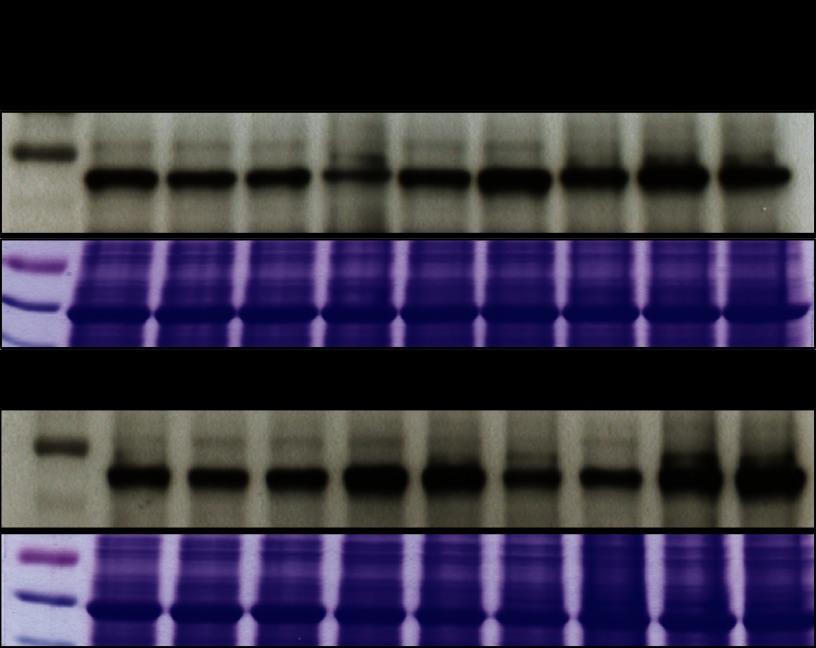
Fig. 4.
SNX1-GFP and mutant expression level. Western blot with 3-dpg seedling expression level was detected by anti-GFP antibody; Coomassie Brilliant Blue staining is shown as the loading control. Lines with severe lateral root inhibition phenotype in Fig. 3 are marked with asterisk.

