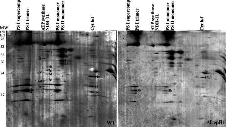
Fig. 2.
Two-dimensional BN/SDS-PAGE separation of membrane protein complexes of WT and ΔLepB1. Protein complexes in WT and ΔLepB1 lanes from the first dimension were separated into their subunits by denaturating 14% SDS-PAGE containing 6 m urea. The numbered protein spots were excised from the Coomassie blue stained gel and identified by LC-MS/MS (See supplemental Data File S1).

