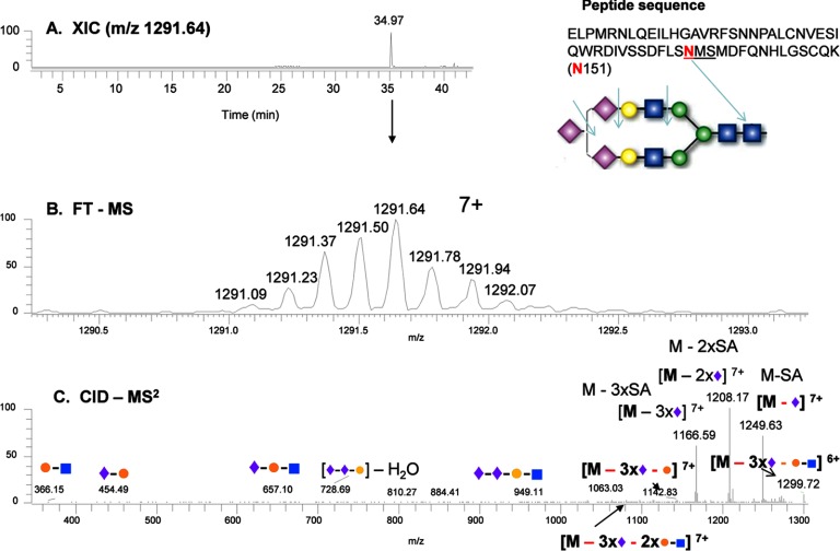
Fig. 2.
Identification of a tri-sialylated glycan at Asn-151 from sEGFR. A, extracted ion chromatogram (XIC) of the glycopeptide containing Asn-151; B, mass and charge of the Lys-C-digested peptide with the anticipated glycan structure; and C, CID-MS2 spectrum of the precursor ion from B. In the glycan structures, the green circle represents mannose; the yellow circle represents galactose; the blue square represents N-acetylglucosamine; the red triangle represents fucose, and the purple diamond represents sialic acid.

