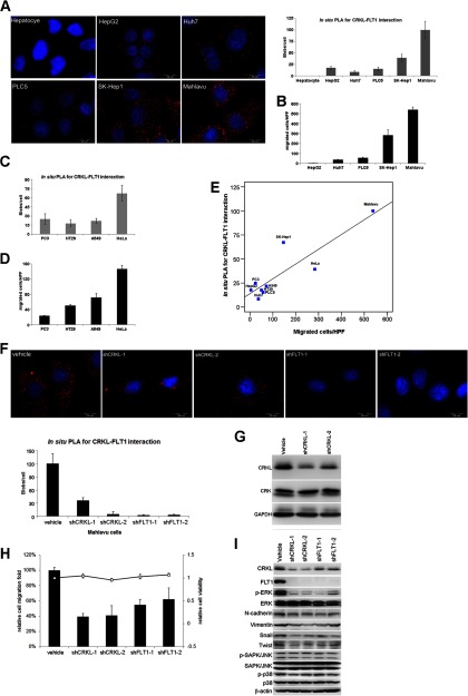
Fig. 3.
Characterization of the CRKL-FLT1 interaction in HCC cell lines. A, images (left) and quantification (right) of in situ PLA signal for CRKL-FLT1 interaction in hepatocytes and five HCC cell lines (HepG2, Huh7, PLC5, SK-Hep1, and Mahlavu) are shown. B, migratory ability and CRKL-FLT1 interaction were evaluated in five HCC cell lines. The Mahlavu cell is highly migratory cells among five HCC cell lines. The CRKL-FLT1 interaction via in situ PLA (C) and migratory ability (D) was measured in other cancers, including prostate cancer cells (PC3), colon cancer cells (HT29), lung cancer cells (A549), and cervical cancer (HeLa). E, CRKL-FLT1 expression was correlated with cell migration. The intensity of in situ PLA for CRKL-FLT1 interaction and migrated cells/high power field showed a positive correlation with a correlation coefficient = 0.886 (p < 0.001) as analyzed for Pearson product-moment correlation when we observed all nine cancer cells. F, five different stable clones of CRKL and FLT1 Mahlavu cells (vehicle (scramble control), shCRKL-1, shCRKL-2, shFLT1–1, and shFLT1–2) with knockdown of CRKL or FLT1 were established. The expression of CRKL-FLT1 interaction was decreased in Mahlavu stable clones with knockdown of CRKL or FLT1 compared with vehicle control cells. G, expression of CRK was not affected by knockdown of CRKL in Mahlavu cells. The shCRKL-1 and shCRKL-2 specifically target CRKL, but not CRK. H, knockdown of CRKL or FLT1 reduced migration ability compared with vehicle control cells. I, lysates from Mahlavu cells with CRKL or FLT1 knockdown were subject to Western blotting for mesenchymal markers and several markers of signaling pathways. Western blot analysis showed that both CRKL and FLT1 participated in the ERK but not in JNK or p38 signaling pathway and that they were also involved in EMT process. Vehicle, scramble control.

