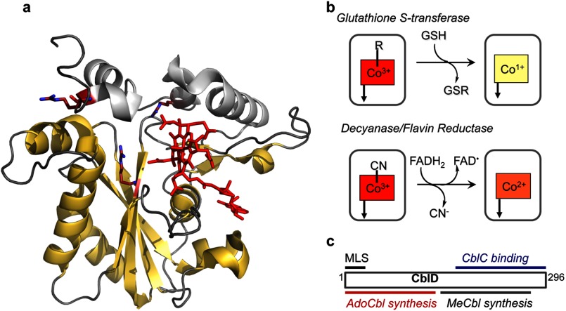
FIGURE 3.
Biochemical functions of CblC and CblD. a, the structure of human CblC with MeCbl (Protein Data Bank code 3SC0). MeCbl (red) is bound in a base-off conformation, with the DMB tail located in a side pocket. The flavin reductase domain is shown in yellow. The arginine residues that are mutated in patients are shown in stick representation. b, reactions catalyzed by CblC. c, localization of mutations in CblD that lead to impaired AdoCbl or MeCbl synthesis and the minimal length required for binding to CblC. MLS, mitochondrial leader sequence.

