FIGURE 4.
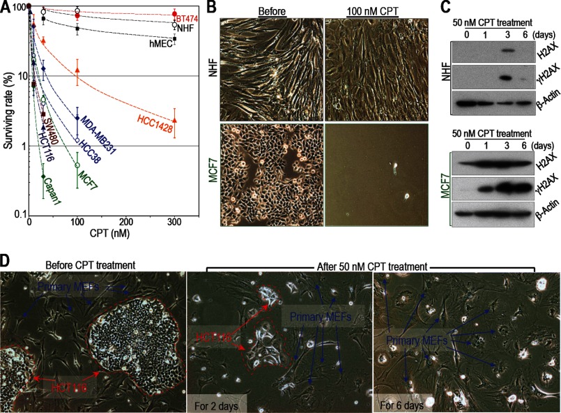
Cancer cells are more sensitive to CPT than normal cells unless they acquire resistance. A–C, cancer cells are generally sensitive to CPT unless they acquire resistance. Cancer cells are more sensitive to CPT than primary NIHs and human mammary epithelial cells (hMEC). However, BT474 breast cancer cells are resistant (A). Sensitivity to damage was determined as outlined in Fig. 1. Survival rates were plotted as in Fig. 1A. B, representative images of NHF and MCF7 cells are shown before and after CPT treatment (other cell lines are shown in supplemental Fig. S3, A and B). C, H2AX and γH2AX levels in MCF7 and NHF cells (levels in other cell lines are shown in supplemental Fig. S3, C and D). The cancer cells used in these experiments harbor mutations in either Arf (HCT116 and MCF7) or p53 (BT474, HCC1428, MDA-MB231, Capan1, HCC 38, and SW480). D, selective killing of cancer cells was examined in a mixed culture of primary WT MEFs and HCT116 (colon cancer) cells. The different cell types are easily distinguished on the basis of morphology and size. HCT116 cells are circled with red dashed lines. Primary WT MEFs are indicated by blue arrows. The images show cells before and after treatment with 50 nm CPT for 2 or 6 days. Both cell types initially showed the flattened and enlarged morphology characteristic of senescent cells (after 2 days). After 6 days, the cancer cells died, but the primary WT MEFs survived.

