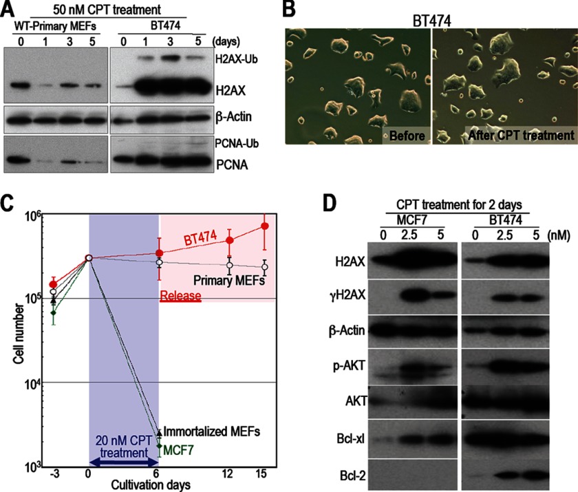
FIGURE 5.
The mechanism underlying the resistance of BT474 cells to CPT is different from that underlying the survival of normal cells. A, the regulatory mechanisms in cancer cells that are resistant to CPT are different from those in surviving normal cells. Responses to 50 nm CPT are shown. Unlike primary WT MEFs, BT474 cells (which are resistant to CPT) accumulated H2AX and maintained their PCNA levels. Ub-H2AX and Ub-PCNA indicate ubiquitinated molecules. B, unlike primary WT MEFs, BT474 cells show morphological changes after CPT treatment. C, each cell type was treated with 20 nm CPT for 6 days, during which most of the immortalized WT MEFs and MCF7 cells died. However, BT474 cells and primary WT MEFs survived. Although BT474 cells recovered growth activity immediately after release from CPT, primary WT MEFs remained quiescent. D, compared with cancer cells that have not developed drug resistance (MCF7 cells), BT474 cells show increased levels of phosphorylated AKT, Bcl-xl, and Bcl2 (all of which promote cell survival).

