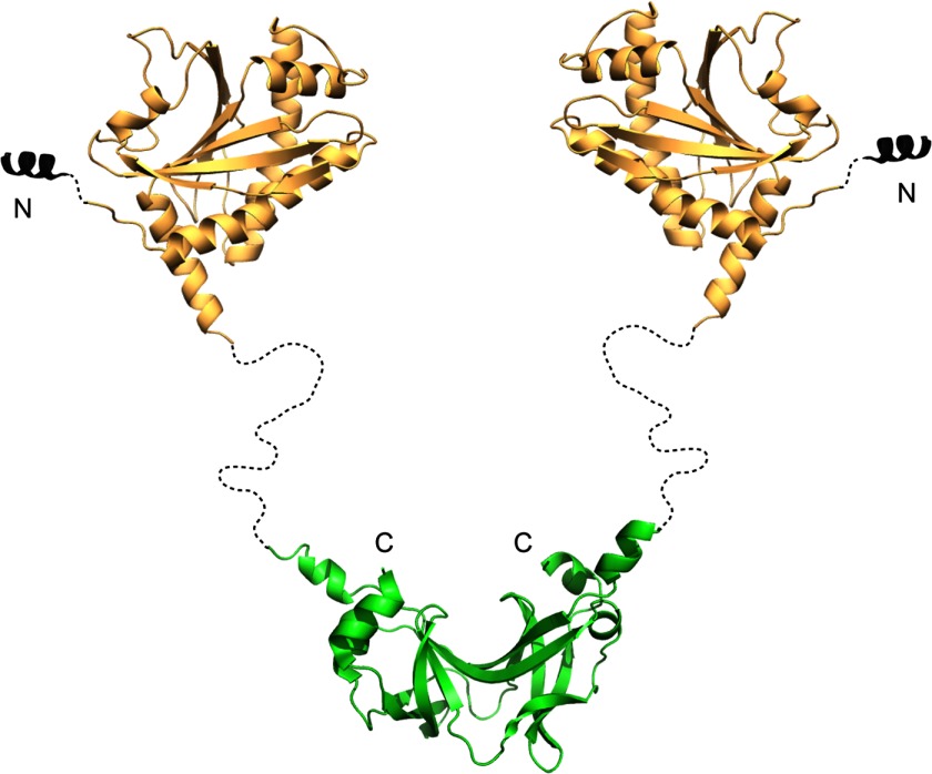
FIGURE 6.
Model of FliY. Model derives from crystal structures of the middle domain (Protein Data Bank code 4HYN) and the C-terminal ∼100 residues (Protein Data Bank code 1YAB) Secondary structure prediction suggests that the long linker region is unstructured. The color pattern as defined in Fig. 1.

