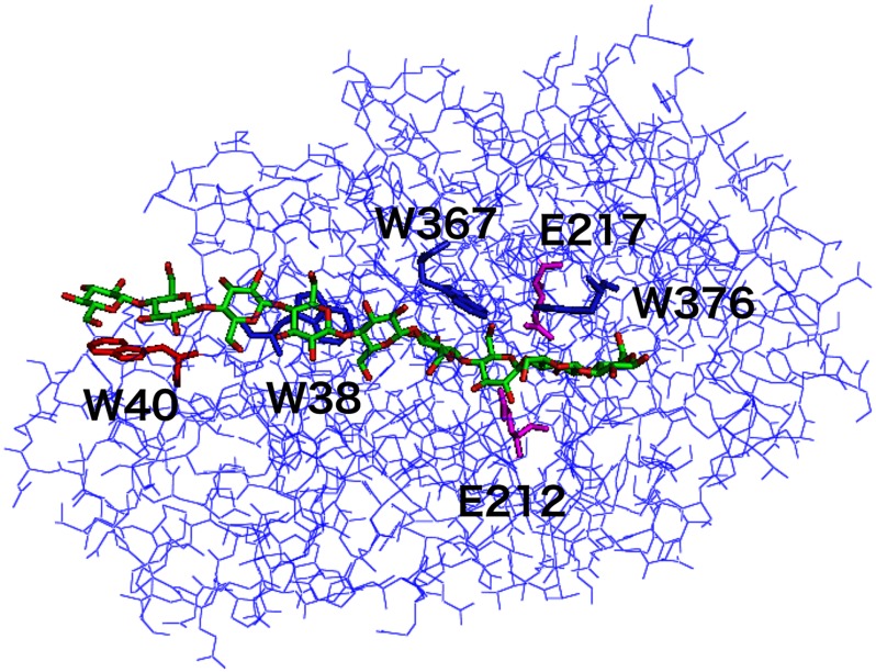
FIGURE 1.
Three-dimensional structure of the catalytic domain of TrCel7A cellobiohydrolase. The α-carbon chain structure of the TrCel7A catalytic domain is shown, together with a modeled continuous cellononaose (Glc-9) chain in the active site tunnel (Protein Data Bank code 8CEL). Trp-40 at subsite −7 is shown in red, and Glu-212, which acts as the catalytic nucleophile between subsites −1 and +1 and was used as a reference position in the molecular dynamics simulations, is shown in magenta.

