FIGURE 3.
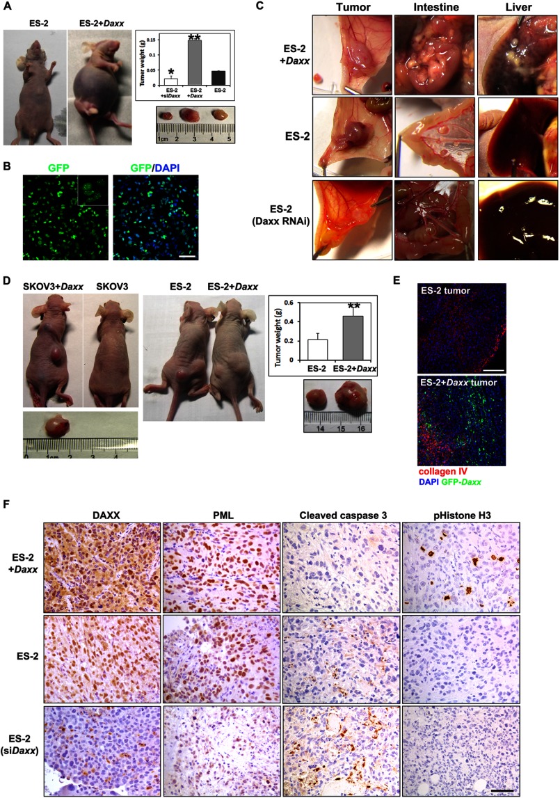
DAXX promotes ovarian cancer cell proliferation and metastasis in vivo. A, ES-2 cells and their derivatives (GFP-Daxx overexpression and siDaxx stable cell lines) were injected intraperitoneally into nude mice (106 cells/mouse). After 30 days, mice that received Daxx-overexpressing ES-2 cells had significant ascites accumulation. Tumors were removed and weighed (n = 18). Results are the means ± S.E. of six independent experiments. B, ascites fluid cells were harvested from nude mice and cultured in vitro. Abundant GFP signals were observed by fluorescent microscopy. Left panel, GFP. Right panel, merge (GFP and DAPI). Scale bar, 50 μm. C, ES-2 cells and their derivatives (GFP-Daxx overexpression and siDaxx stable cell lines) were injected underneath the abdomen membrane of nude mice (106 cells/mouse). In situ colonization of tumor cells and metastasis to the intestine and liver were examined 3 weeks later. D, ovarian cancer cells (SKOV3 and ES-2) with or without DAXX overexpression (106 cells for each) were implanted subcutaneously into different nude mice, respectively. After 30 days, tumors were removed and weighed (n = 10). E, cryosections were prepared from tumor tissues derived from ES-2 and DAXX-overexpressing ES-2 cells. Prominent angiogenesis was shown by immunofluorescent staining for collagen IV (red). DAXX overexpression was shown by GFP fluorescence (green). Tissues were counterstained with DAPI (blue). Scale bar, 100 μm. F, immunohistochemistry staining for DAXX, PML, cleaved caspase3, and pHistone H3 in representative ES-2-derived tumor tissues. Scale bar, 50 μm.

