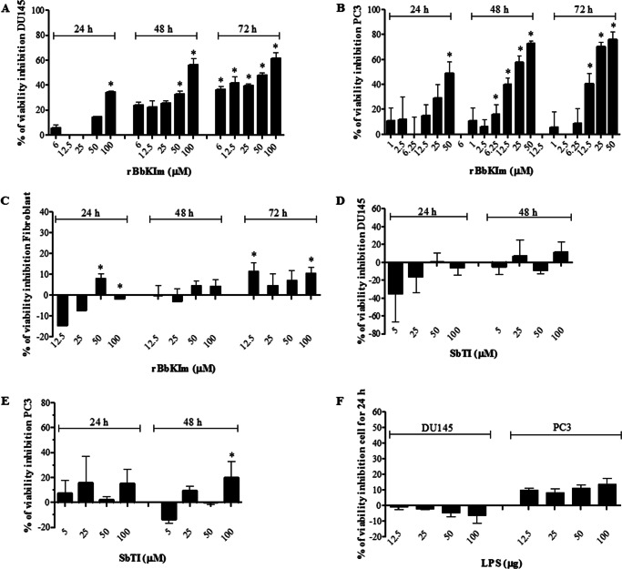
FIGURE 1.
The effect of rBbKIm, SbTI, and LPS on cell viability. A–C, DU145 (A), PC3 (B), and fibroblast (C) cells were treated with increasing concentrations (0–100 μm) of rBbKIm for 24, 48, and 72 h. D and E, DU145 (D) and PC3 (E) cells were treated with increasing concentrations (0–100 μm) of SbTI for 24 and 48 h. F, DU145 and PC3 cells were treated with increasing concentrations (0–100 μm) of LPS for 24 h. Cell viability was measured using the MTT assay. The control cells were treated with a medium containing 7 mm HEPES, pH 7.4 (vehicle). The calculation of the percentage of inhibition was performed for the control cells (untreated) at each incubation time. The values are expressed as the means ± standard deviation of the representative experiment. Significant differences versus controls are presented (ANOVA; *, p < 0.05).

