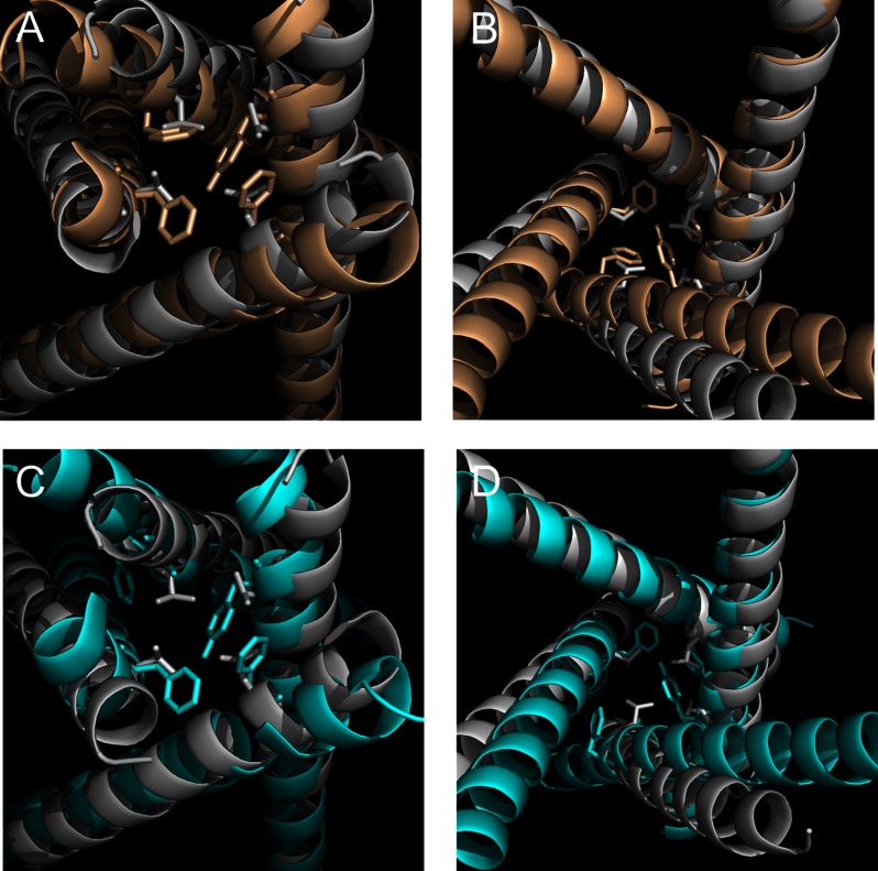
FIGURE 5.
Alignment of NaV1.7 WT and Del-L955 mutant channel structural model with NaVRh structures. A, cytoslic view of the alignment of NaV1.7 structural model with that of NaVRh. NaV1.7 is shown in wheat, and NaVRh is shown in gray. Tyr405 of DI, Phe960 and Ser961 of DII, Phe1449 of DIII, and Phe1752 of DIV are shown in stick configuration. Activation gate of NaVRh (Leu219) is also shown in stick configuration. B, extracellular view of the alignment of NaV1.7 structural model with NaVRh. C, cytoslic view of the alignment of Del-L955 mutant channel structural model with NaVRh. Del-L955 is shown in cyan, and NaVRh is shown in gray. D, extracellular view of the alignment of Del-L955 mutant channel structural model with NaVRh.

