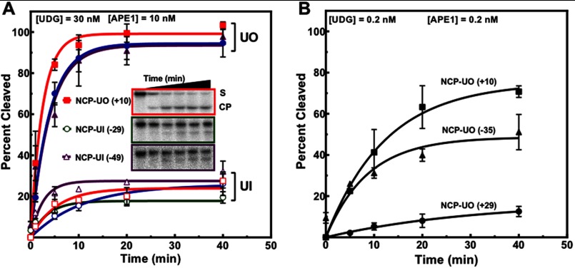
FIGURE 2.
Assessment of the removal of rotationally and translationally positioned uracils by UDG and APE1. A, NCPs containing a single uracil at different sites were incubated with UDG and APE1. Open symbols represent in uracils as follows: red square, NCP-UI (+4) and open blue circle, NCP-UI (+25). Filled symbols correspond to out uracils as follows: solid blue circle, NCP-UO (+29) and purple triangle, NCP-UO (−35). B, NCPs containing uracil with DNA backbones outwardly oriented were treated with low equimolar enzyme concentrations of 0.2 nm. Data points represent the mean ± 1 S.D. of at least three independent experiments and were fitted to a single phase exponential curve as described under “Experimental Procedures.” For data points where the error bars are not visible, the standard deviations were smaller or similar in magnitude to the size of the symbols.

