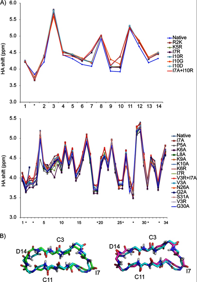
FIGURE 3.
A, comparison of the secondary shifts of selected SFTI-1 and MCoTI-II mutants. The shifts were generated by subtracting the αH shifts from random coil shifts (50). B, alignment of NMR structures of SFTI-1 backbone with the SFTI-1 variants I10R (left) and R2A (right). The parent peptide SFTI-1 is displayed in pale cyan. The variants I10R and R02A are displayed in green and purple respectively.

