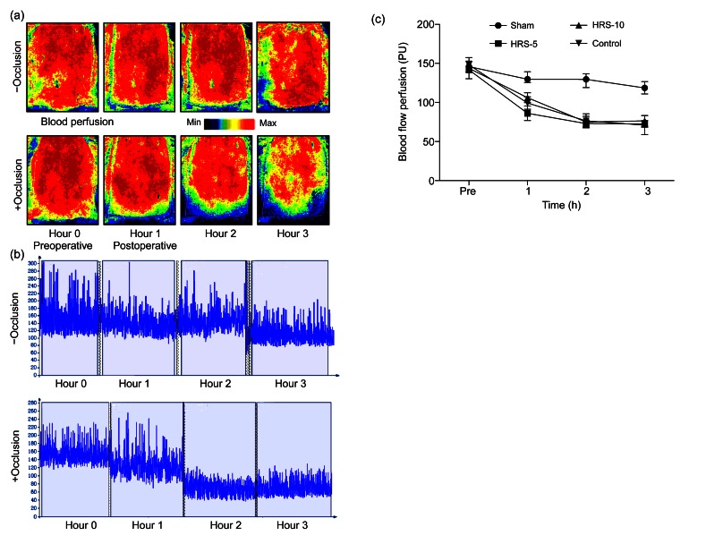
Fig. 4.
Laser speckle contrast imaging for measuring changes in blood flow perfusion during the perioperative period
(a) Laser speckle contrast imaging was used to estimate perfusion before the operation and at 1, 2 and 3 h after the operation. The region of interest (ROI) was the entire abdominal flap. Occlusion for 3 h was enough to cause skin flap ischemia. The color scale illustrates variation in blood flow from maximal (red) to minimal (dark) perfusion. (b) Flux patterns revealed that occlusion successfully caused the decrease in perfusion from 150 PU to 70 PU, whereas the sham group without occlusion had a relatively stable blood perfusion. The image acquisition rate was 3 s−1 and lasted for about 3 min at Hour 0, Hour 1, Hour 2, and Hour 3. (c) There was no significant difference in blood perfusion between the four groups before the operation. The sham group had a much higher perfusion rate at the 3 h time point. The results are expressed as mean±SEM (n=15) (Note: for interpretation of the references to color in this figure legend, the reader is referred to the web version of this article)

