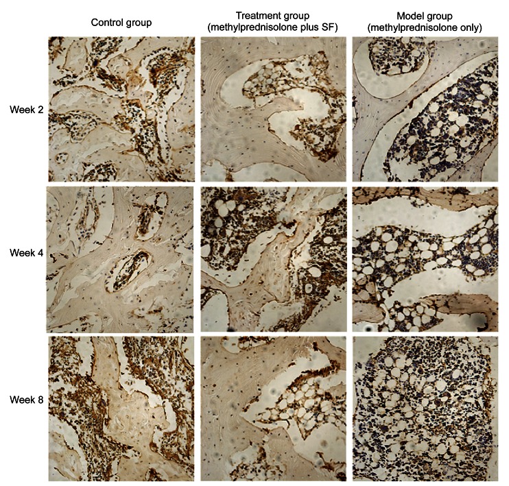
Fig. 7.
Effect of methylprednisolone and methylprednisolone plus sodium ferulate (SF) on osteocytes in immunohistochemical staining of Bcl-2 at Weeks 2, 4, and 8
Week 2: The positive staining is brown, and the staining in the model group was less intense than that in the control and treatment groups. The visible positive staining was hardly observed in the model group. Week 4: The positive staining was most significant in the treatment group, and lowest in the model group. Week 8: The positive staining in the treatment group was intense and similar with that in the control group. The positive staining in the model group slightly increased but was far less than that in the other two groups. Magnification: ×200 (Note: for interpretation of the references to color in this figure legend, the reader is referred to the web version of this article)

