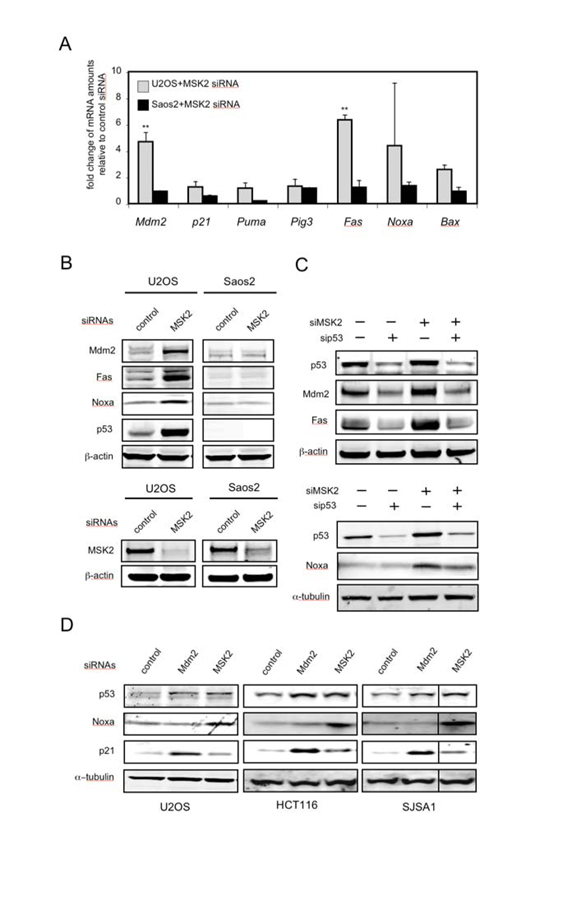
Fig. 2.
Knock-down of MSK2 results in selective activation of p53 target genes. (A) Induction of endogenous p53 targets by transfection of the indicated cells with MSK2 siRNA. The mRNA abundance of the indicated genes was analyzed 72 hours after transfection by quantitative real-time PCR (qRT-PCR). Error bars represent SDM from at least three samples assayed in duplicate. The Ct values were corrected by β-actin and fold expression values are relative to control siRNA. Statistical significance was determined by ANOVA and calculating the p values for the comparisons between cell lines. * P < 0.05; ** P < 0.01. (B) p53-containing U2OS cells and p53-deficient Saos2 cells were transfected with the indicated siRNAs. Protein abundance was determined 72 hours later by Western blotting (top). Detection of endogenous MSK2 was possible only after immunoprecipitation. Identical amounts of total protein were immunoprecipitated with an MSK2 antibody and then analyzed by Western blot using a different MSK2 antibody. β-actin amounts in the inputs are shown (bottom). (C) U2OS cells were transfected first with control or p53 siRNAs and were transfected 24 hours later with control, Mdm2, or MSK2 siRNAs. Protein abundance was analyzed 48 hours after the second transfection. (D) p53-positive U2OS, HCT116, and SJSA1 cells were transfected with the indicated siRNAs. Protein abundance was analyzed 72 hours later by Western blotting.

