
Fig 5.
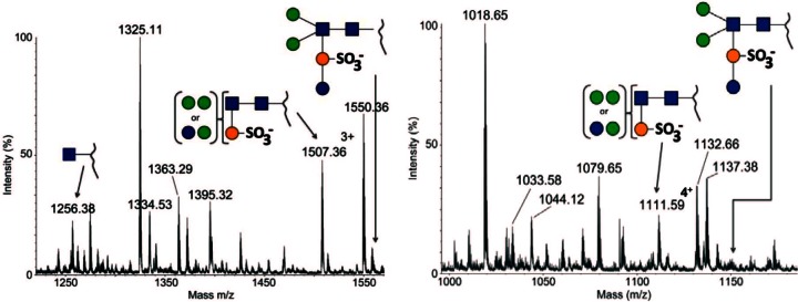
Detailed analysis of the S. acidocaldarius Δagl16 mutant background S-layer glycopeptide. The total ion chromatograms of the glycan profiles for the tryptic peptides T24 (A) and T26 (B) are shown. The annotated peaks show that the truncated Hex2QuiS1HexNAc2 glycoform is present on the observed glycopeptides, whereas the full glycan is missing. For reference, a cartoon depicting the full glycan is provided for each spectrum. Glycan symbols: blue sphere, glucose; green sphere, mannose; orange sphere, sulfoquinovose; blue square, GlcNAc.