
Fig 7.
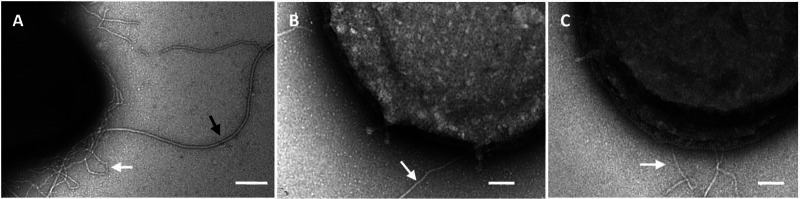
TEM analysis of negatively stained cells of MW001 (A), the Δagl3 mutant (B), and the Δagl16 mutant (C) showed the lack of archaella on each of the agl deletion mutants. The MW001 background strain exhibits Aap pili (8 to 10 nm; white arrow) as well as the archaella (10 to 14 nm, black arrow); in the agl deletion mutants, only the Aap pili could be detected. Bars, 200 nm (A) and 100 nm (B and C).