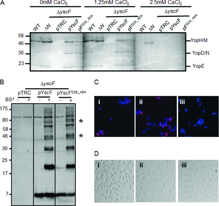
Fig 3.
Characterization of the YscFD28AD46A needle in Y. pseudotuberculosis. (A) The WT IP2666, ΔyopN, or ΔyscF strain carrying pTRC99A (pTRC), pTRC99A-yscF (pYscF), or pTRC99A-yscFD28AD46A (pFD28_46A) was grown in 2× YT medium plus 5 mM EGTA and 20 mM MgCl2 supplemented with the indicated amounts of CaCl2 for 2 h at 26°C. Expression of YscF from pTRC99A was induced with the addition of IPTG when bacteria were shifted to 37°C. Supernatants from cultures grown for 2 h at 37°C were precipitated with TCA. Secreted proteins were separated by SDS-PAGE and visualized by Coomassie blue. (B) Bacteria were grown under secretion-inducing conditions for 90 min and then exposed to 1 mM BS3 or water for 30 min. Whole cells were solubilized in sample buffer and subjected to Western blot analysis with antibody against YscF. Asterisks indicate HMW bands that vary in intensity between the WT YscF- and YscFD28AD46A-expressing strains. pYscFD28_46A, pTRC99A-yscFD28AD46A. (C) Y. pseudotuberculosis cells were grown under secretion-inducing conditions for 1.5 h and then fixed and mounted onto coverslips. Cells were labeled with anti-YscF antibody and visualized with Alexa Fluor-594-conjugated anti-rabbit secondary (red). Bacteria were counterstained with DAPI (blue). Frame i, ΔyscF(pTRC99A-yscF); frame ii, ΔyscF (pTRC99A-yscFD28AD46A); frame iii, ΔyscF(pTRC99A). (D) HEp-2 cells were seeded into 96-well plates and then infected with Y. pseudotuberculosis at an MOI of 50:1. Images were taken after 1 h of incubation at 37°C. Frame i, ΔyscF(pTRC99A-yscF); frame ii, ΔyscF(pTRC99A-yscFD28AD46A); frame iii, ΔyscF(pTRC99A). Each experiment was repeated a minimum of two times. Representative experiments are shown for each.

