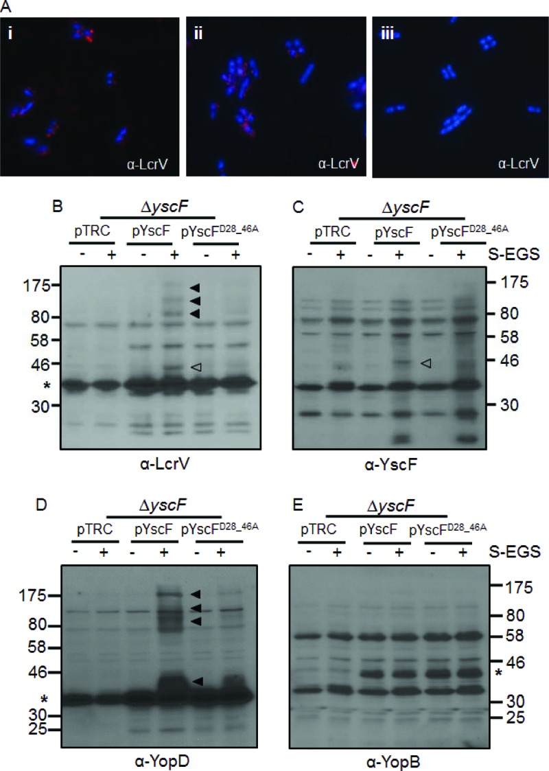
Fig 4.
LcrV cross-linked complexes are altered on Y. pseudotuberculosis expressing YscFD28AD46A polymers. (A) Y. pseudotuberculosis cells were grown under secretion-inducing conditions for 1.5 h, fixed, mounted onto coverslips, and labeled with anti-LcrV antibody and then with Alexa Fluor-594-conjugated anti-rabbit secondary antibody (red). Frame i, ΔyscF(pTRC99A-yscF); frame ii, ΔyscF(pTRC99A-yscFD28AD46A); frame iii, ΔyscF(pTRC99A). (B to E) ΔyscF strains expressing pTRC99A, pTRC99A-yscF, or pTRC99A-yscFD28AD46A were grown in secretion medium at 37°C. Expression of YscF was induced with the addition of 30 μM IPTG followed by incubation at 37°C. Cross-linking with sulfo-EGS (S-EGS) was performed as described in the legend for Fig. 1. Proteins were detected with anti-LcrV (B), anti-YscF (C), anti-YopD (D), or anti-YopB (E) antibodies. Open arrowheads indicate the YscF-LcrV complex; filled arrowheads indicate HMW cross-linked bands. Asterisks indicate the monomeric protein; YscF is not visible on the gel. The experiment was repeated twice, and a representative blot is shown.

