
Fig 5.
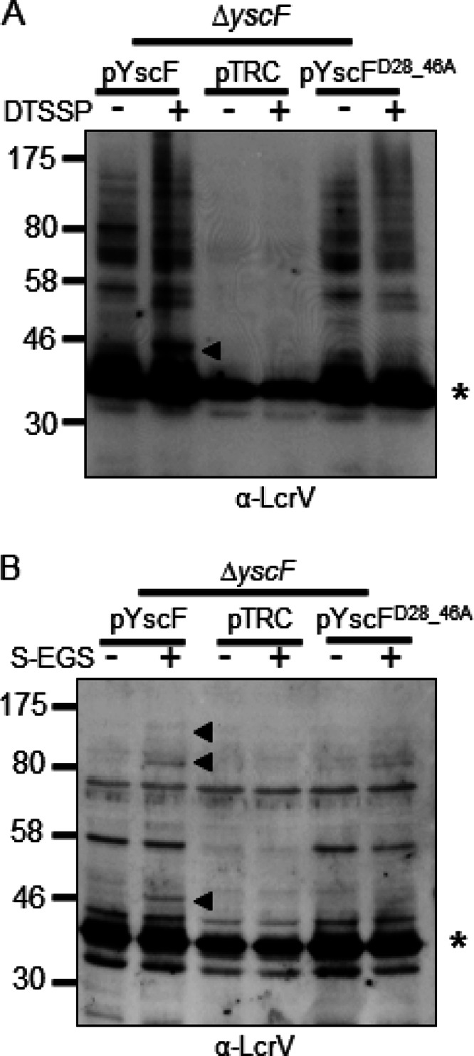
YscF-LcrV and HMW LcrV complexes from a YscFD28AD46A strain are poorly detected with DTSSP or when grown in minimal medium. (A) A ΔyscF strain expressing pTRC99A (pTRC), pTRC99A-yscF (pYscF), or pTRC99A-yscFD28AD46A (pYscFD28_46A) was grown in secretion medium at 37°C. Expression from pTRC99A was induced with the addition of 30 μM IPTG followed by incubation at 37°C for 1.5 h. Cells were incubated with 1 mM DTSSP or water for 30 min at 37°C. Bacteria were solubilized, and proteins were analyzed by Western blotting with anti-LcrV antibody. (B) Strains expressing pTRC99A, pTRC99A-yscF, or pTRC99A-yscFD28AD46A were grown in M9 minimal medium supplemented with 0.4% glucose and 1% defined amino acid mix lacking arginine, glutamine, lysine, and asparagine. Bacteria were incubated for 2 h at 26°C; 30 μM IPTG was added, and bacteria were grown at 37°C for 3 h. Cultures were exposed to 1 mM sulfo-EGS for 30 min at 37°C. Proteins were analyzed by Western blot analysis. Arrows indicate YscF-LcrV and HMW complexes formed by the addition of cross-linking agents. Asterisks indicate monomeric LcrV. Each experiment was repeated twice and a representative blots are shown.