
Fig 6.
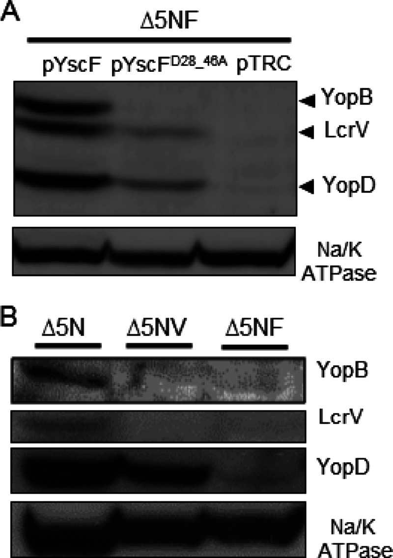
Association of LcrV, YopB, and YopD with HEp-2 plasma membranes after infection with the WT YscF or YscFD28AD46A strain. HEp-2 cells were infected with Y. pseudotuberculosis strains at an MOI of 50:1. After 1 h, the plasma membranes were collected, and the proteins were separated by SDS-PAGE. Proteins were visualized by Western blot analysis with anti-YopB, anti-LcrV, anti-YopD, and anti-Na/K ATPase antibodies. (A) Strains lacking yopHEMOJN yscF (Δ5NF) and expressing pYscF, pYscFD28AD46A, or pTRC99A were induced in 30 μM IPTG at 37°C. (B) Western blot analysis of the ΔyopHEMOJN (Δ5N), ΔyopHEMOJN ΔlcrV (Δ5NV), and ΔyopHEMOJN ΔyscF (Δ5NF) strains. The experiments were performed twice, and a representative blot is shown.