
Fig 7.
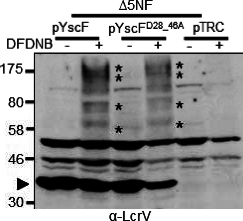
Membrane-associated LcrV forms oligomers in both WT YscF- and YscFD28AD46A-expressing strains. Y. pseudotuberculosis ΔyopHEMOJN ΔyscF (Δ5NF) expressing pTRC99A, pTRC99A-yscF, or pTRC99A-yscFD28AD46A was grown at 37°C for 1.5 h with 30 μM IPTG to induce expression of YscF. The bacteria were then used to infect HEp-2 cells at an MOI of 50:1. After a 1-h incubation at 37°C, HEp-2 cells were collected, and the bacteria were removed by centrifugation. Lysates were incubated with 500 μM DFDNB before plasma membrane vesicles were isolated. Proteins were separated by SDS-PAGE and analyzed by Western blotting with anti-LcrV antibody. Asterisks indicate HMW LcrV complexes formed by the addition of DFDNB. The experiment was repeated twice, and a representative blot is shown.