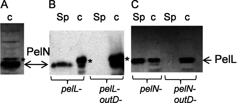
Fig 3.

Secretion of the PelN protein by the Out system. After separation of the proteins by SDS-PAGE, PelL antibodies were used to detect the proteins PelN and PelL by Western blotting. (A) The periplasmic fraction was prepared from IPTG-induced cells of E. coli BL21(DE3)/PNA13 overproducing PelN. Cultures of various D. dadantii mutants were used to prepare culture supernatants (Sp) and cell lysates (c). (B and C) PelN was detected in the pelL and pelL-outD mutants (B), and PelL was detected in the pelN and pelN-outD mutants (C). In each D. dadantii strain, the pecS gene was also inactivated in order to increase PelL and PelN production. Although the development usually lasted 1 min to detect PelL, it was increased to 10 min for PelN detection in D. dadantii. A nonspecific band is indicated with an asterisk.
