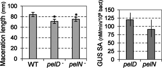
Fig 6.

Infection of chicory leaves with the pelN mutant. (A) The length of macerated tissue was measured after a 24 h infection with the mutant and with the wild-type strain 3937 (WT). (B) The β-glucuronidase (GUS) assay and bacterial enumeration were performed on the macerated tissue in order to estimate the expression of the uidA fusions in pelD and pelN. The mean value and the standard deviations reported correspond to 10 infections with each strain. Asterisks indicate statistically significant differences in the degree of maceration of the mutants, compared to the wild-type strain (P < 0.05 [Student t test]).
