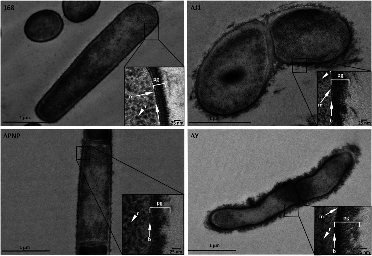
Fig 5.
RNase Y and RNase J1 mutants have altered cell walls. Shown are representative transmission electron microscopy images of the wild type (WT) and the pnp (ΔPNP), rnjA (ΔJ1), and rny (ΔY) mutants. The peptidoglycan (pg) layer, an electron-dense layer representing the base (b) of the peptidoglycan layer, the thin (white) cellular membrane (m), and ribosomes (r) are indicated where visible.

