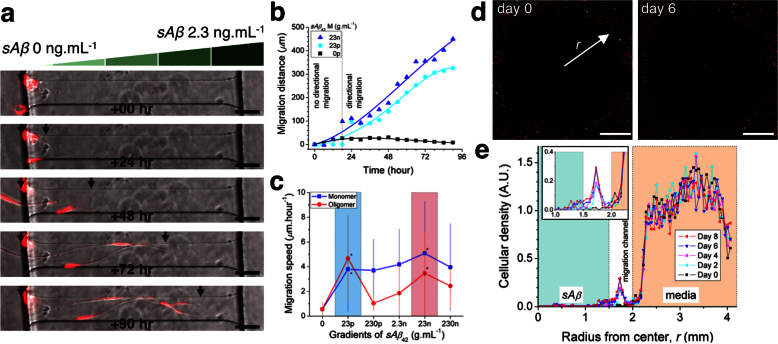
Figure 2. Inducement of microglial directional migration by gradients of soluble Aβ.
(a) Individual microglia migrate directionally along the gradient of sAβ monomers formed in migration channels. (b) Activation of directional motility can be discerned after 24 hours-exposure to gradients of sAβ42 in monomers at 23 pg.mL−1 and 23 ng.mL−1. (c) Dose-dependence of microglia migration speed during 4 days observations reveal two peak activities under gradients of soluble Aβ42 monomers and oligomers at concentrations of 23 pg.mL−1 and 23 ng.mL−1 (ranges highlighted in cyan and pink colors, respectively). (d) Fluorescence images present detectable microglia accumulation toward the source of soluble Aβ42 at day 6 compared to day 0. (e) Microglial density profiles at different days quantify the migration of microglia populations toward the source of soluble Aβ42 in monomers at 2.3 ng.mL−1. See the details on the stability and the preparation of soluble monomeric and oligomeric Aβ in Figs. S1, S2, and Supplementary methods. (Student's t-test. * P < 0.01 with respect to no sAβ42). ncell = 18 for each condition. Data represent mean ± s.e.m.

