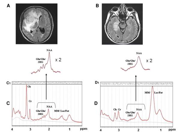
Fig. 1.
Magnetic Resonance Imaging and MRS. Top panel (A and B): Axial MRI scans of two anaplastic astrocytomas (WHO grade III) show focal regions of fluid-attenuated inversion recovery (FLAIR) hyperintensity corresponding to areas of tumor. Other than the surrounding edema related to the size of the tumors, pre-operative structural MRI imaging characteristics were generally indistinguishable between IDH1 mutant (A) and wild-type (B) gliomas of the same grade and histopathology. Voxels of interest for MR spectroscopic analysis were localized by two neuroradiologists. Spectral voxels were placed in the center of the area of solid tumor, excluding regions of probable necrosis or vasogenic edema. Comparison of representative MR spectra from IDH1 mutant gliomas (C) versus wild-type spectra (D). Note the extra peaks in the region of Glu/Gln/2-HG (centered at 2.25 ppm) that are increased in the IDH1mutant tumors, compared to the wild-type MR spectra

