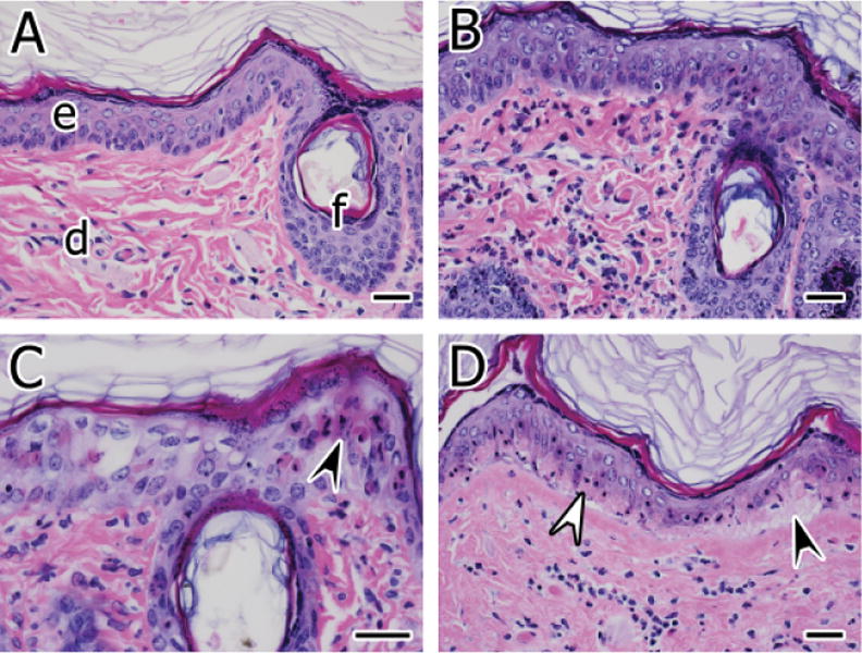
Figure 1.

(A) Normal skin from an air-exposed control site. The dermis (d) contains a few scattered, mostly mononuclear leukocytes. The epidermis (e) and a follicular infundibulum (f) are also indicated. (B) Skin from a site exposed to sulfur mustard (SM) for 6 minutes at 6 hours postexposure. Inflammatory cells composed of heterophils and mononuclear leukocytes are scattered in the superficial dermis. Some of the superficial acanthocytes are mildly swollen and nuclei are vesicular (intracellular edema). (C) Epidermal epithelial apoptosis (arrowhead) and intracellular edema are progressively more severe at 24 hours postexposure to SM for 6 minutes. (D) Basal epithelial necrosis characterized by hypereosinophilic cytoplasm, nuclear pyknosis, and karyorrhexis is present in the epidermis (white arrowhead) at 24 hours postexposure to SM for 12 minutes. Most of the acanthocytes superficial to the basal cells retain viable features. The affected epidermis is minimally separated from the underlying dermis (black arrowhead). Hematoxylin and eosin stain, bars = 30 μm.
