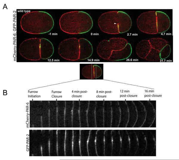
Fig. 1. Dynamics of PAR-6-mCherry and PAR-2-GFP proteins during cell division.
(A) Embryos expressing both PAR-6-mCherry and PAR-2-GFP were imaged using spinning disk confocal microscopy from one minute prior to furrow formation through the four-cell stage. Localization of PAR-6-mCherry and PAR-2-GFP are temporally and spatially localized along the cleavage furrow membrane. PAR-6 and PAR-2 localize to the furrow membrane during ingression but are distinct in that PAR-2-GFP is excluded from the extreme tip of the furrow (white arrowhead, Fig. 1A, 2.7 min), where PAR-6-mCherry is only found. Once the midbody has formed, PAR-2-GFP becomes restricted to the midbody region (the midbody membrane accumulation plus the membrane flanking it) postfurrow closure (Fig 1A, 12.5–14.9 min and Fig 1B). During the next division the localization of PAR-2-GFP disappears from the midbody region and localizes to the entire cortex of the P2 blastomere (Fig 1A, 26.6–31.7 min). At this time, PAR-6-mCherry is now found at the cortex and adjoining membranes of the anterior blastomeres (ABa, ABp, and EMS) at the four-cell stage (Fig 1A, 31.7 min). The adjoining membrane adjacent to the P2 blastomere in EMS and ABp is labeled with PAR-2-GFP. (B) View of PAR-6-mCherry and PAR-2-GFP dynamics along the cleavage furrow at the mid-focal plane. Corresponding red and green channels were separated and a montage of furrow ingression was built from frames 80 s apart from furrow initiation to 16-min post-closure. At 4-min postclosure, PAR-6 and PAR-2 are evenly distributed along the furrow membrane and accumulate at the midbody. After the 4-min postclosure, PAR-6-mCherry remains evenly distributed along the furrow membrane whereas, PAR-2-GFP becomes restricted to the midbody (a bolus of membrane) and regions flanking the midbody by the end of the time course.

