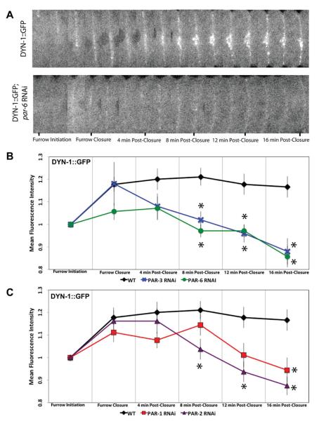
Fig. 4. Anterior and posterior PAR proteins influence DYN-1 localization to the furrow.
Single, mid-focal plane confocal images of the cell equator from time-lapse sequences of embryos expressing DYN-1-GFP. (A) Montages show a control embryo and an embryo in which PAR-6 was depleted. DYN-1-GFP is maintained along the furrow throughout cytokinesis and 16-min postfurrow closure concentrating at the midbody. In PAR-6 RNAi-treated embryos, a reduction in recruitment of DYN-1-GFP to the cleavage furrow is observed. (B) Analysis of DYN-1-GFP intensity at the furrow in wild-type and anterior PAR (PAR-3 and PAR-6) depleted embryos. (C) Analysis of DYN-1-GFP fluorescence intensity at the cleavage furrow in posterior PAR depleted embryos. Depletion of both anterior and posterior PAR proteins leads to a decrease in fluorescence intensity of DYN-1-GFP at the cleavage furrow. The number of embryos for each experiment is listed as: wild type, n = 9, par-1 RNAi n = 9, par-2 RNAi n = 8, par-3 RNAi n = 5, par-6 RNAi n = 7. Asterisks indicate time points that displayed a significant difference (A P-value of <0.05) between corresponding PAR depletion and wild type fluorescence intensity values. Statistical significance was determined by ANOVA followed by post hoc testing.

