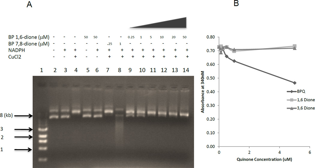
Figure 7.
DNA strand scission and NADPH consumption by PAH quinones (A) p53 cDNA plasmid was incubated with vehicle (DMSO) alone, (lane 1); 0.25 µM and 1 µM B[a]P-7,8-dione in the presence of 10 mM NADPH and 100 µM CuCl2 for 2h at 37 °C (lanes 7 and 8); 0.25 µM, 1 µM, 5 µM, 10 µM, 20 µM and 50 µM B[a]P-1,6-dione in the presence of 10 mM NADPH and 100 µM CuCl2 for 2h at 37 °C (lanes 9–14). The treated p53 was directly analyzed by electrophoresis on a 1% agarose gel. (B) NADPH oxidation was analyzed by absorbance at 340 nm.

